Axonal loss in the multiple sclerosis spinal cord revisited.
Brain Pathol. 2017 Apr 12. doi: 10.1111/bpa.12516. [Epub ahead of print]
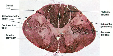
Preventing chronic disease deterioration is an unmet need in people with multiple sclerosis, where axonal loss is considered a key substrate of disability. Clinically, chronic multiple sclerosis often presents as progressive myelopathy. Spinal cord cross-sectional area assessed using MRI predicts increasing disability and has, by inference, been proposed as an indirect index of axonal degeneration. However, the association between cross-sectional area and axonal loss, and their correlation with demyelination, have never been systematically investigated using human post mortem tissue. We extensively sampled spinal cords of seven women and six men with multiple sclerosis (mean disease duration= 29 years) and five healthy controls to quantify axonal density and its association with demyelination and cross-sectional area. 396 tissue blocks were embedded in paraffin and immuno-stained for myelin basic protein and phosphorylated neurofilaments. Measurements included total cross-sectional area, areas of (i) lateral cortico-spinal tracts, (ii) grey matter, (iii) white matter, (iv) demyelination, and the number of axons within the lateral cortico-spinal tracts. Linear mixed models were used to analyse relationships. In multiple sclerosis cross-sectional area reduction at cervical, thoracic and lumbar levels ranged between 19 and 24% with white (19-24%) and grey (17-21%) matter atrophy contributing equally across levels. Axonal density in multiple sclerosis was lower by 57-62% across all levels and affected all fibres regardless of diameter. Demyelination affected 24-48% of the grey matter, most extensively at the thoracic level, and 11-13% of the white matter, with no significant differences across levels. Disease duration was associated with reduced axonal density, however not with any area index. Significant association was detected between focal demyelination and decreased axonal density. In conclusion, over nearly 30 years multiple sclerosis reduces axonal density by 60% throughout the spinal cord. Spinal cord cross sectional area, reduced by about 20%, appears to be a poor predictor of axonal density.
In this study we looked at the amount of nerve content in the spinal cord and found signfcant loss in people with long standing MS. They people were in wheelchairs at the time they passed and they had lost 60% of their nerve content.
However when we measured the spinal cord area it did not shrink by that much despite a large amount of nerve loss.
As this is a central measure, detected by MRI used to suggest nerve loss, it suggests that MRI measures of atrophy are missing an awful lot.
Whilst there may be alot of nerve loss the space can be filled by other cells or fluid. This may be why we see pseudoatrophy with drugs that block inflammation, this removes swelling and so the nerve looks like it shrinks. Therefore, one of the central measures of progression is probably flawed.
CoI: This work of TeamG & S
